The Modeling Approach
Myo- and fibroblast activity is a critical factor in closing the wound and achieving the best scar outcome after burn injury. TGF-beta1 is the primary driver of fibroblast-to-myofibroblast differentiation and subsequent collagen I synthesis (Deng et al., Signal Transduct Target Ther, 2024), while the balance between pro- and anti-inflammatory signals at the phase transition determines whether the wound resolves toward normotrophic scarring or pathological fibrosis. The MechaProlif model focuses on this transition from the inflammatory to the proliferative phase and into early remodeling, capturing the key cell types and molecular signals that govern this window.
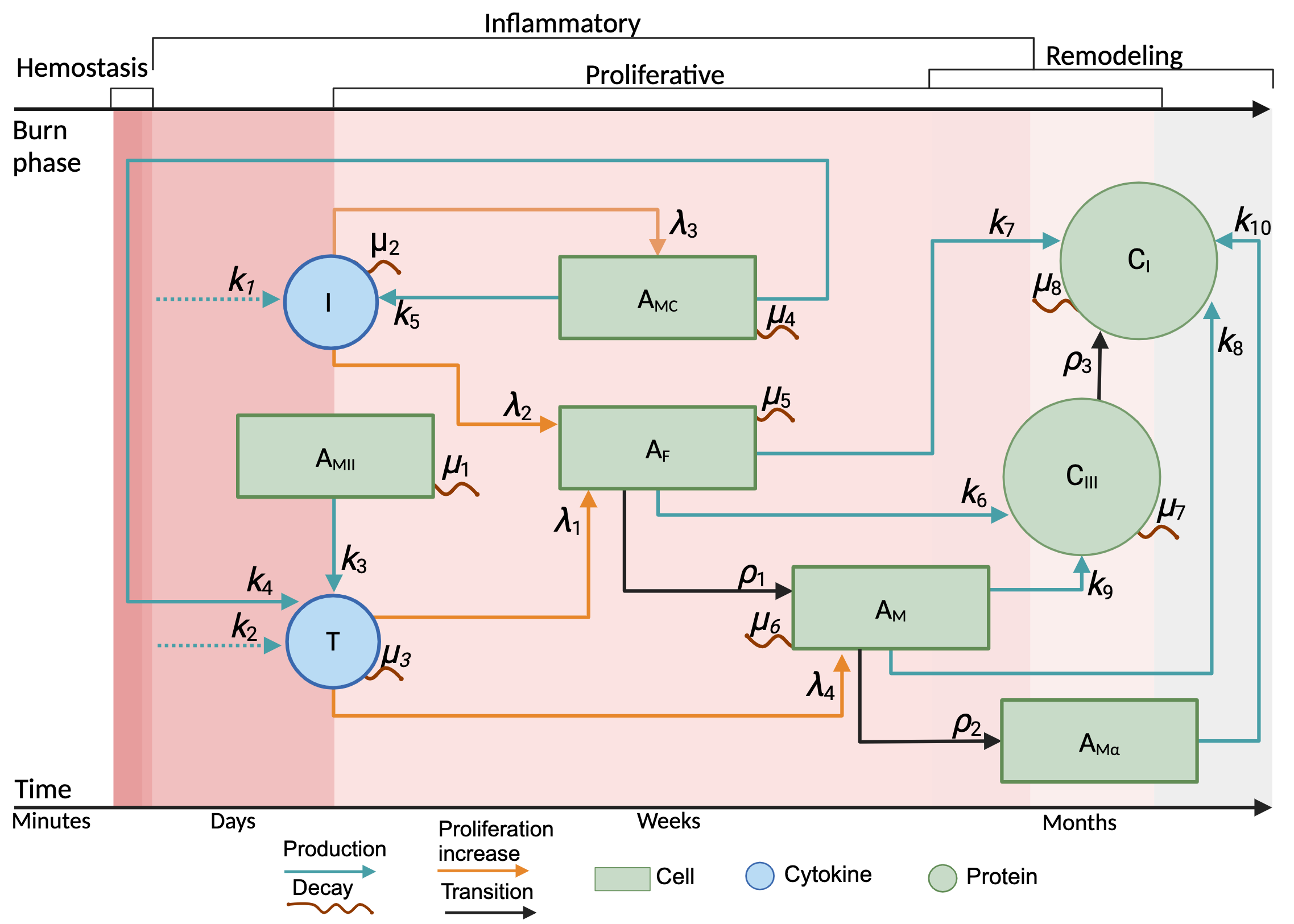
Rate parameters: κ (production/proliferation increase), λ (transition rates), ρ (decay rates), and μ govern the dynamics of cell populations and protein concentrations.
The model revolves around the end of the inflammatory phase and the beginning of the proliferative phase, using equilibrium values from the inflammatory model as initial conditions.
TGF-β1 is the primary driver of myofibroblast differentiation and collagen I synthesis. IL-8 mediates neutrophil recruitment and modulates the transition between phases.
Simulation Scenarios
Two distinct inflammatory profiles were tested to understand how variations in the transition from inflammation to proliferation affect cell dynamics and collagen deposition:
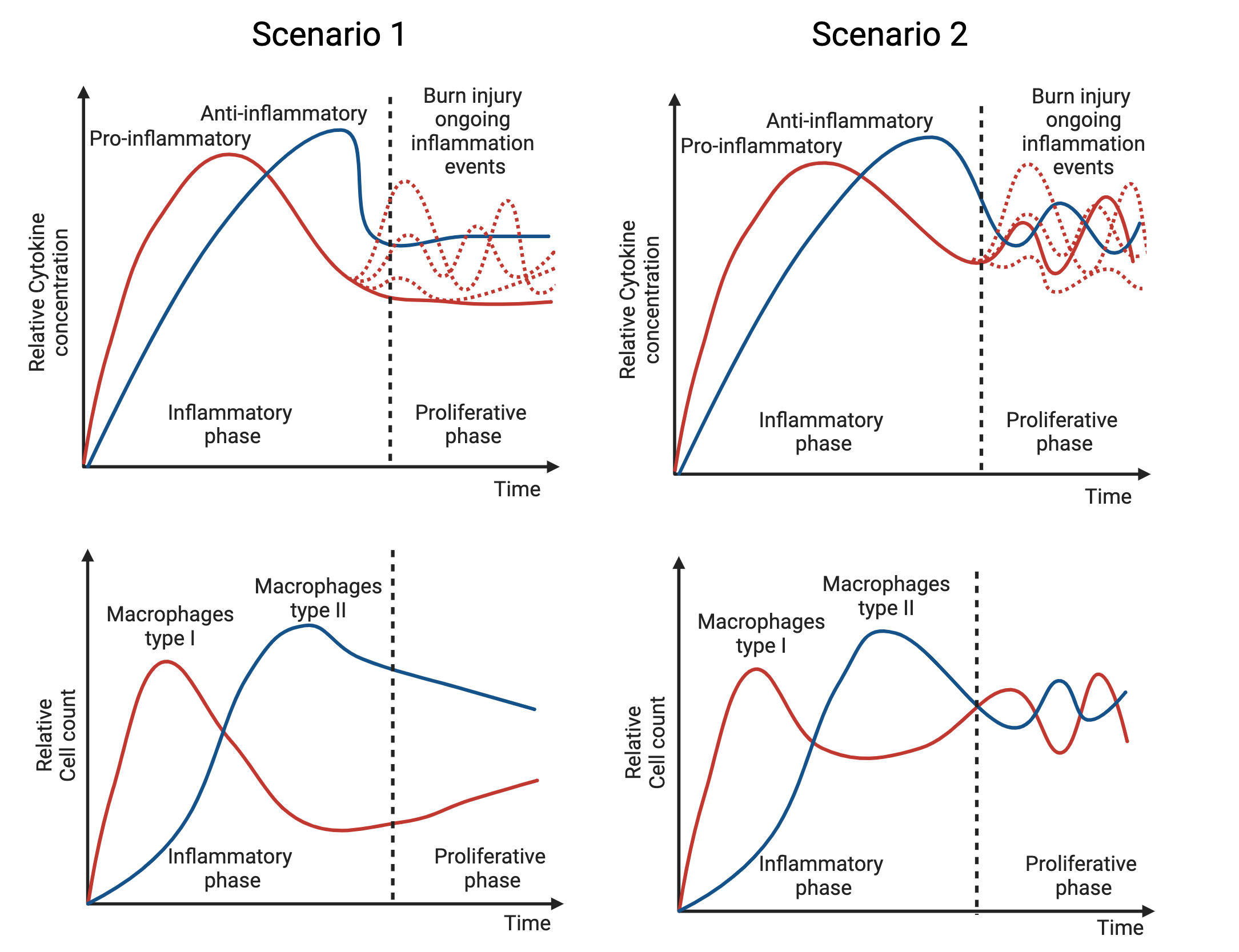
The dotted black line in each plot marks the assumed starting conditions for the proliferative phase, derived from inflammatory phase equilibrium values (Figure 1).
Parameter search was conducted to fit dynamics consistent with what is observed in burn wounds as a first simulation. Full results in the IEEE publication.
Anti-inflammatory dominance with periodic pro-inflammatory bursts
The inflammatory stage ends with PI cytokines near AI levels, but regular intense inflammatory episodes, triggered by continuous stimuli, periodically drive PI concentrations much higher. Designed to analyse the effect of recurrent inflammation on proliferative cell activity: does periodic PI increase amplify or suppress wound closure?
Ambiguous interplay between pro- and anti-inflammatory signals
PI and AI mediators are intertwined during the phase transition, with neither clearly dominant. Event-driven inflammation can significantly spike PI signals, complicating pattern identification. Macrophage type I/II balance is unspecified, challenging accepted polarisation models and their role in wound healing dynamics.
How to Read the Model
In Figure 1, the network diagram shows the relationships between all agents and proteins from the beginning of the proliferative phase. The dotted black line in Figure 2 shows expected dynamics for each cell type and cytokine at phase entry. These serve as initial conditions.
From those values, the model propagates the interactions described in Figure 1. The parameter search fits dynamics that are biologically plausible and relatable to what actually occurs in burn wounds. For full results, consult the IEEE publication.
Simulator: Coming Soon
An interactive parameter simulator for the MechaProlif model is currently in development. It will allow direct exploration of how changing κ, λ, ρ, and μ affects wound closure dynamics and collagen composition.